
Objective
Many complex fluids are heterogeneous on a µm scale. This is especially true for biological sys-tems like the cytoskeletal protein filament networks, which provide stability and mobility of euka-ryotic cells, but also for polymers from biological or synthetic resources, which are used e.g. as thickeners in food, personal and health care, plant protection or coating and adhesive formula-tions. Video Particle Tracking (VPT) is a microrheological tool, which allows us to study such micro-structural and micro-mechanical heterogeneities, since it gives direct access to the trajec-tories and mean square displacements of embedded tracer particles subject to thermal motion. Even living cells can be studied with high spatial resolution. But also bulk rheological properties can be extracted even for heterogeneous samples, when cross-correlations of displacements of particles which are far apart (~10-100 µm) are analyzed (so-called two-point microrheology).
Operating Principle
We use a standard set-up for particle tracking video microscopy including an inverted fluores-cence microscope and a CCD camera for image acquisition of fluorescent molecules or colloi-dal tracer particles. This set-up gives access to a wide range of timescales, from high-speed video rate to unbounded long time-lapse acquisitions. Roughly a hundred tracer particles can be tracked simultaneously and the ensemble averaged mean-squared displacement (MSD) is ex-tracted while still retaining each of the individual particle trajectories. Commercially available algorithms are used for automatic identification of particle centers with sub-pixel accuracy. Once a particle is located in a sequence of video images, particle positions in each image are corre-lated with positions in later images to calculate trajectories.
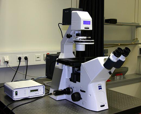
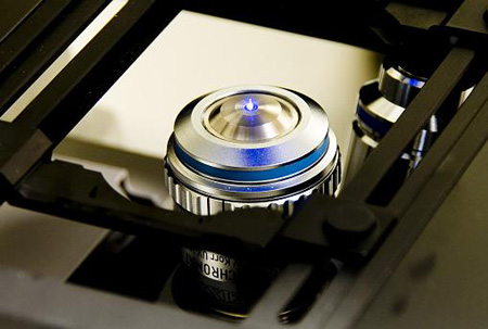
Fig. 1: Inverted fluorescence microscope AxioObserver D, Zeiss (left); C-Apochromat 40x Objective, 1.2 numerical aperture, water immersion (right).
Specifications
| Microscope: | Inverted fluorescence microscope (AxioObserver D, Zeiss), LED light source (Colibri, Zeiss) for illumination. |
| Objective: | Fluar 100x, 1.3 numerical aperture, oil-immersion lens; C-Apochromat 40x, 1.2 numerical aperture, water-immersion lens. |
| Camera: | sCMOS camera Zyla X (Andor Technology), 50 fps, 2560 x 2160 px |
| Tracer: | Fluorescent (green) micro-spheres: P(S/A/V-COOH) 0.13, 0.21, 0.51, 0.9 μm (Bangs Laboratories). |
| Tracking: | Image Processing System, Visiometrics iPS. Additionally self-written MatLab program, based on Crocker and Grier algorithm. |
Applications
- Cells and biological fluids, e.g. DNA, myosin, F-actin, ...
- Polymer solutions
- Surfactant solutions
- Suspensions and emulsions
Literature
- Mason, T. G., and D. A. Weitz. 1995. Optical measurements of frequency dependent linear viscoelastic moduli of complex fluids. Phys. Rev. Lett. 74:1250–1253
- Chen, D. T., E. R. Weeks, J. C. Crocker, M. F. Islam, R. Verma, J. Gruber, A. J. Levine, T. C. Lubensky, and A. G. Yodh. 2003. Rheological microscopy: local mechanical properties from microrheology. Phys. Rev. Lett. 90:108301
- Apgar, J., Y. Tseng, E. Fedorov, M. B. Herwig, S. C. Almo, and D. Wirtz. 2000. Multiple-particle tracking measurements of heterogeneities in solutions of actin filaments and actin bundles. Biophys. J. 79:1095–1106
- Valentine, M. T., P. D. Kaplan, D. Thota, J. C. Crocker, T. Gisler, R. K. Prud’homme, M. Beck, and D. A. Weitz. 2001. Investigating the microenvironments of inhomogeneous soft materials with multiple particle tracking. Phys. Rev. E. 64:061506